Figure 4.
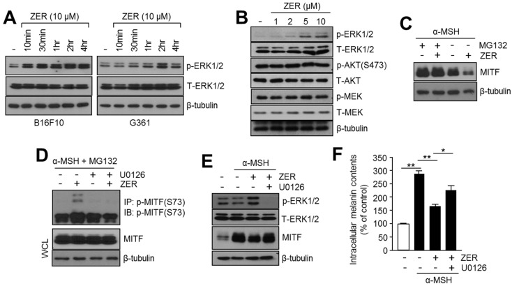
Activation of ERK1/2 is involved in the anti-melanogenic effect of zerumbone (ZER). (A) Phosphorylation of ERK1/2 upon ZER treatment. B16F10 and G361 cells were incubated with 10 μM ZER for different time intervals as indicated. Protein expression levels of p-ERK1/2, total-ERK1/2, and β-tubulin (loading control) were analyzed by immunoblotting; (B) Alterations in signaling pathways upon ZER treatment. B16F10 cells were treated with different concentrations of ZER for 1 h, and then the total protein was analyzed by immunoblotting to determine activation of ERK1/2, AKT, and MEK as described in the “Materials and Methods” section; (C) Proteasome inhibitor, MG132, blocks ZER-mediated reduction of MITF. B16F10 cells were pre-treated with 10 μM ZER for 1 h, and then cells were incubated in the absence (−) or presence (+) of MG132 (20 μM) for 6 h; (D) ZER increases phosphorylation of MITF. B16F10 cells were pre-treated with 20 μM MG132 for 3 h, and then cells were incubated in the absence (−) or presence (+) of ZER (10 μM) and U0126 (10 μM) for 1 h. Phosphorylated MITF was determined by immunoprecipitation and immunoblotting; (E) Immunoblot analysis of MITF and p-ERK1/2 in absence (−) or presence (+) of ZER or U0126, a selective inhibitor of MAPK. B16F10 cells were treated with U0126 (10 μM) and ZER (10 μM), and were subsequently incubated with α-MSH (0.1 mM) for 1 h (for p-ERK1/2 and T-ERK1/2 detection) and 4 h (for MITF and β-tubulin detection); and, (F) Estimation of intracellular melanin content in absence and presence of U0126 and ZER, in response to α-MSH stimulation. Cells were cultured with ZER (10 μM) and U0126 (10 μM), and incubated with α-MSH (0.1 mM) for three days, as indicated. Detailed procedure for melanin content analysis is described in “Materials and Methods” section. Values represent mean ± SD of three independent experiments performed in triplicate; * p < 0.05 and ** p < 0.01.

