Figure 1.
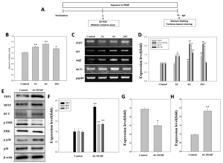
Effect of pulsed electromagnetic fields (PEMFs) on the pigmentation of zebrafish after exposure to PEMFs. (A) Schematic representation of the schedule of the zebrafish pigmentation study. After PEMF exposure, the melanin content was determined. (B) The melanin content in zebrafish (n = 80) was detected using the melanin content assay at 5 dpf. (C) The mRNA expression, detected using reverse transcription polymerase chain reaction, results in zebrafish (n = 20) after exposure to PEMFs at 5 dpf. (D) The mRNA expression of melanogenesis-related genes, using glyceraldehyde-3-phosphate dehydrogenase (GAPDH) as the reference gene. The effect of PEMF on the protein levels of tyrosinase-related protein-1 (TRP1), microphthalmia-associated transcription factor (MITF), dopachrome tautomerase (DCT), extracellular signal-regulated kinase (ERK), p-ERK, p-p38, p38, and b-actin in zebrafish (n = 30) at 15 dpf. (E) The Western blotting analysis of the pigmentation-related proteins; (F) TRP1, MITF, and DCT expression level; (G) p-ERK expression; (H) p-p38 expression. Each bar represents the mean ± standard error of the independent experiments performed in triplicate (n = 5). * p < 0.05, ** p < 0.01, compared to the control.

