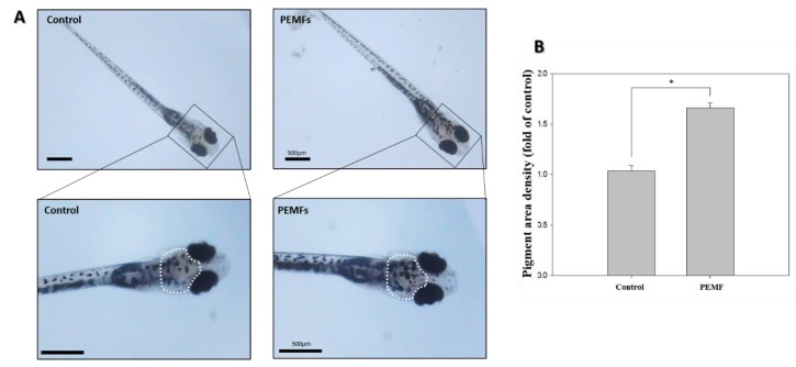
Figure 2.
Effects of 4 G PEMF on pigmentation in zebrafish. (A) Synchronized embryos (n = 20) were exposed to the PEMF at the indicated intensity and frequency. The effects on zebrafish pigmentation were observed under a stereomicroscope via inferior views of the embryos at 15 dpf. (B) The pigmentation area density in the treated embryos, indicated by a white outline, was normalized to that of the control embryos using the ImageJ software. * p < 0.05, compared to the control. The scale bar is 500 μm.

