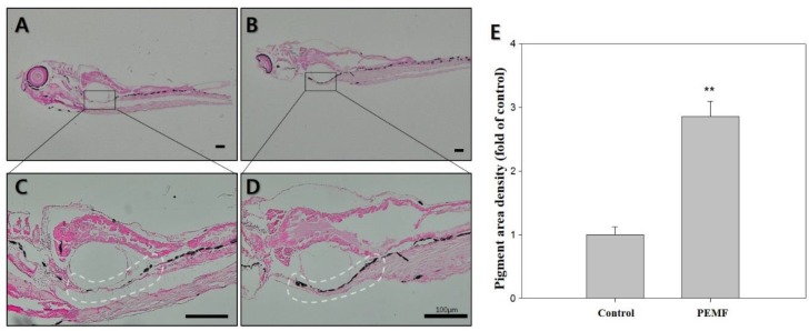
Figure 3.
Effects of 4G PEMF in zebrafish at 15 dpf. Representative images of Fontana-Masson-stained zebrafish (dark color indicates secreted melanin). (A,C) control, (B,D) EMF. (E) the pigmented area density in figure (A–D) was normalized to that of the control, using ImageJ sotware. ** p < 0.05, compared to the control. Original magnification: (A,B) ×40; (C,D) × 100 scale bar; 100 μm.

