
Fig. 4.
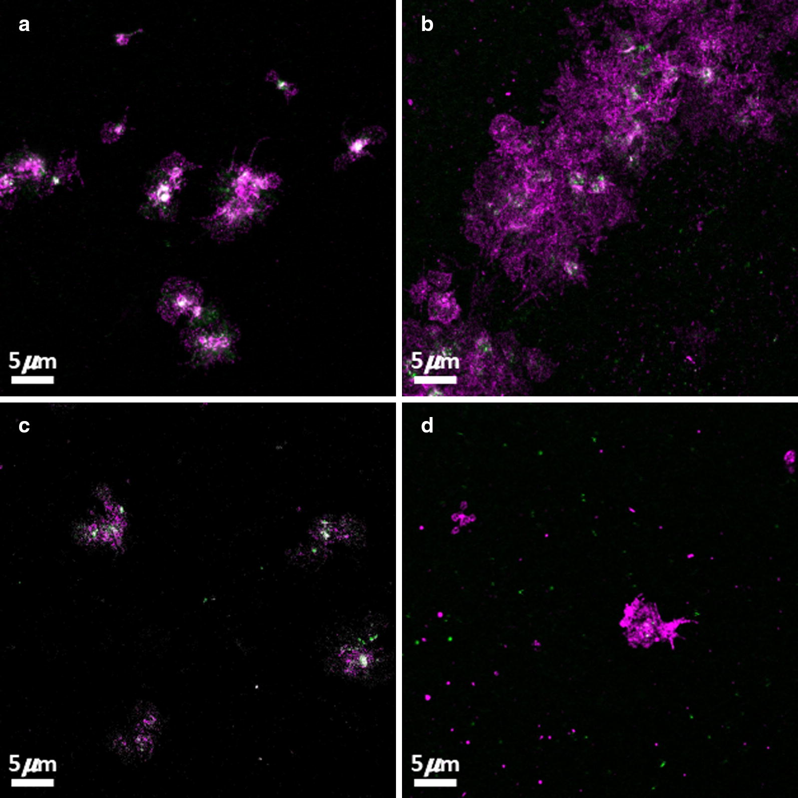
Confocal microscopy where platelets where incubated with CD41 (magenta) and PAC-1 (green). a, c Representative micrograph of platelets from healthy individuals with HbA1c values of 5.0% and 5.2% respectively. Both individuals also reported with CRP levels of < 1.00, indicative of no inflammation. b, d Representative micrographs of platelets from individuals diagnosed with type 2 diabetes mellitus. These individuals had HbA1c levels of 7.0% and 7.2% respectively