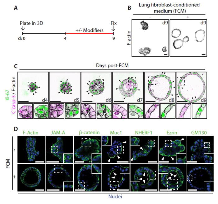
Figure 1. Fibroblast Conditioned Medium (FCM) induces the formation of polarized Calu-3 acini.
(A) Schematic showing the experimental procedure for developing Calu-3 acini in 3D with key modifiers added or removed between days 4–9.
(B) Calu-3 cells were cultured on a layer of Matrigel in standard growth medium containing 2% Matrigel. After 4 days the media on the resultant aggregates were either replenished or replaced with FCM. Acini were fixed on day 9, stained with F-actin and imaged using a confocal microscope.
(C) Acini were fixed and stained at different time points post addition of FCM at day 4, with anti-Ki-67 (green), or anti-Cleaved Caspase-3 (magenta) antibodies and with Phalloidin (F-actin, black). Arrows indicate the localization of Ki-67. Magnified images are also shown (lower panels).
(D) Untreated aggregates or FCM-treated acini were fixed and stained with Hoechst (Nuclei, blue) and either Phalloidin (F-actin), or anti-JAM-A, anti-β-catenin, anti-Muc1 anti-NHERF-1, anti-Ezrin or anti-GM130 antibodies (all green). Arrows indicate the localization of apical proteins. Insets show magnified images.
All scale bars, 50 μm.

