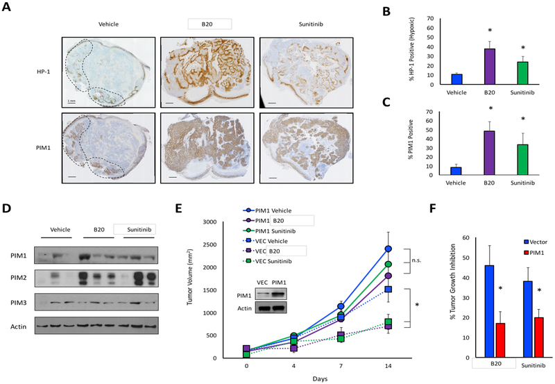
Figure 1. PIM1 expression is increased following treatment with anti-angiogenic agents in vivo.
A) Mice harboring PC3 prostate cancer xenograft tumors were treated with vehicle, B20–4.1.1, or sunitinib. Serial sections from tumors were immunostained to measure hypoxia (hypoxyprobe; HP-1) and PIM1 (dashed lines delineate regions of hypoxia). B) Percent HP-1 and C) percent PIM1 positivity was determined by microscopy. D) PIM isoform expression in each cohort was assessed by immunoblotting. E) Mice injected with PC3/VEC or PC3/PIM1 cells were treated with vehicle, B20–4.1.1, or sunitinib, and tumor volume was measured over time. F) Percent tumor growth inhibition in PC3/VEC and PC3/PIM1 following treatment with sunitinib or B20. *, p < 0.05; n.s. = not significant.

