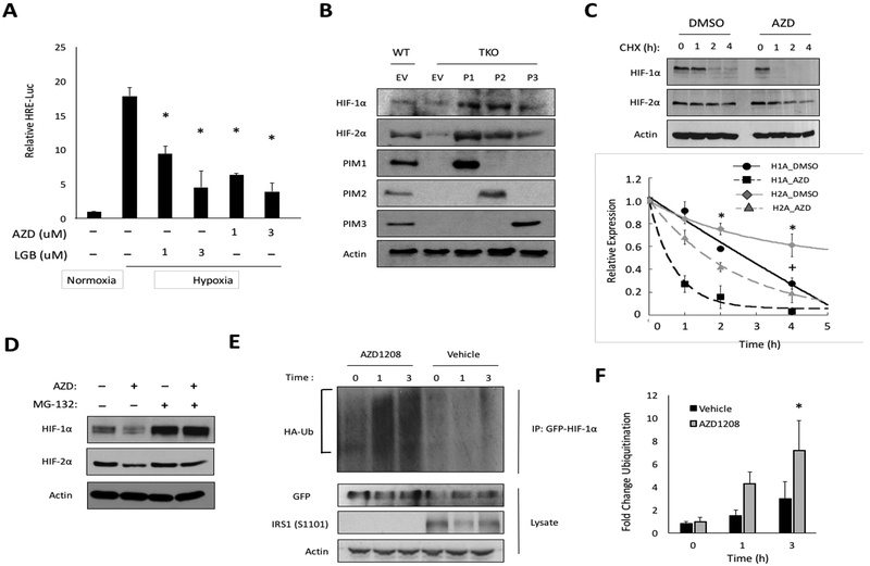
Figure 4. PIM inhibition reduces HIF-1/2α protein levels by enhancing their proteasomal degradation.
A) PC3-LN4 cells expressing HRE-Luc were incubated in normoxia or hypoxia ± AZD1208 or LGB321 for 6 h. B) The indicated cell lines were incubated for 6 h in hypoxia, and protein was collected to monitor HIF1/2-α levels. C) PC3-LN4 cells were cultured in hypoxia for 4 h prior to the addition of cycloheximide (CHX, 12.5 μg/mL). Lysates were harvested at the indicated time points, and densitometry was used to determine the rate of protein decay. D) PC3-LN4 cells were treated with MG-132 (5 μM), AZD1208 (3 μM), or a combination of both for 6 h in hypoxia. E) 293T cells were transfected with GPF-HIF-1α and HA-Ubiquitin and placed in hypoxia for 4 h prior to treatment with AZD1208. Cellular ubiquitination assays were performed, and F) densitometry was used to quantify relative ubiquitination. *, p < 0.05 compared to control.

