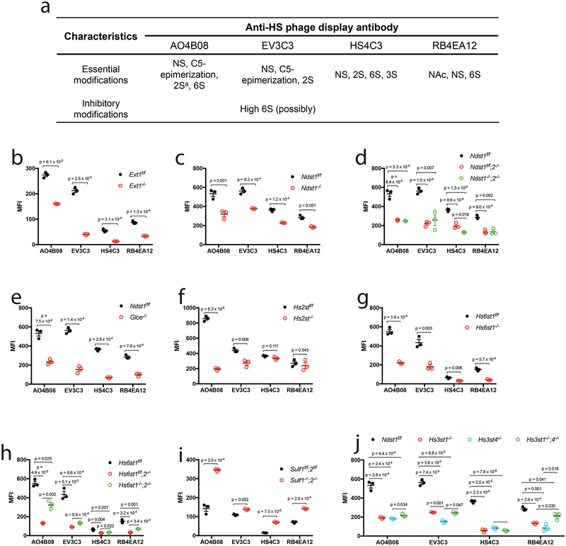
Figure 4. Binding of anti-HS phage display antibody to mutant HS on endothelial cell surface.
a. Summarized characteristics of the HS modifications involved in binding or inhibiting the binding of the anti-HS phage display antibodies in reported biochemical studies. a, antibody AO4B08 requires an internal IdoA2S residue for binding. b-j. Cell surface binding of the antibody. The HS mutant MLECs and their wildtype controls were incubated with the VSV-G-tagged anti-HS phage display antibody, and the cell surface bound antibody was quantified by flow cytometry after further staining the cells with biotinylated anti-VSV-G antibody and fluorescein-tagged streptavidin. The data were summarized from 3 independent experiments and are presented as mean ± SD. Statistical analyses were performed using two-sided, Student`s t-test.

