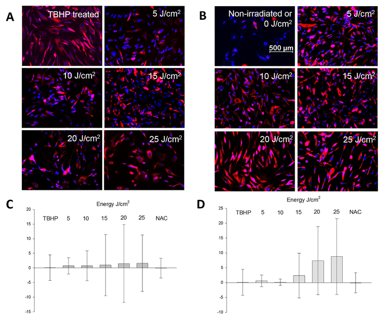
Fig. 3.
Detection of oxidative stress in dermal fibroblasts: Primary cells were treated with 6-carboxy-2′,7′-dichlorodihydrofluorescein diacetate for detecting intracellular Reactive Oxygen Species (ROS) after exposure to lasers of wavelength, λ = 636 nm (A) or λ = 825 nm (B) and fluences ranging in the order of 5 J/cm2. ROS stained orange-red under kaede red and the nuclei stained blue with DAPI filters using Axiovision v4.8 (Zeiss microscopy). Fluorescence intensities were measured for laser wavelength, λ = 636 nm (C) or λ = 825 nm (D) from the digital micrographs using ImageJ (NIH, USA). Tert-Butyl hydroperoxide (TBHP) was the positive control and untreated (0 J/cm2) indicate background of ROS in cells. Scale bar is set as 500 μm across all images. (For interpretation of the references to colour in this figure legend, the reader is referred to the web version of this article.)

