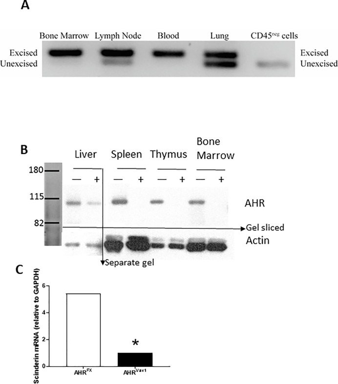
Fig 1. Genotyping and gene induction in 8 week old AHRVav1 mice.
(A) Excised Ahr is detectable in hematopoietic tissues of AHRVav1 animals. Tissues from AHRVav1 and AHRFX mice were isolated as described and DNA was extracted for PCR analysis using a three primer reaction that amplifies either 180 bp fragments (excised allele) or 140 bp (unexcised allele) fragments. (B) AHRVav1 animals have reduced levels of AHR protein in hematopoietic cells. Various tissues (liver, spleen, thymus, and total bone marrow) were analyzed by western blot to detect AHR (~90kD). AHRFX mice (+) express the receptor in all tissues. AHRVav1 (-) mice display reductions or lack of AHR in the tissues examined. Hepatoma cells were used as a positive control as they express large amounts of AHR.(C) Lineage negative cells from AHRVav1 mice fail to upregulate Scinderin mRNA after 6h exposure to 10nM TCDD. Data expressed relative to DMSO vehicle, using HPRT as an endogenous control gene for normalization. N = 4 mice per treatment group. Relative fold change in expression was calculated using the 2ΔΔCt method.

