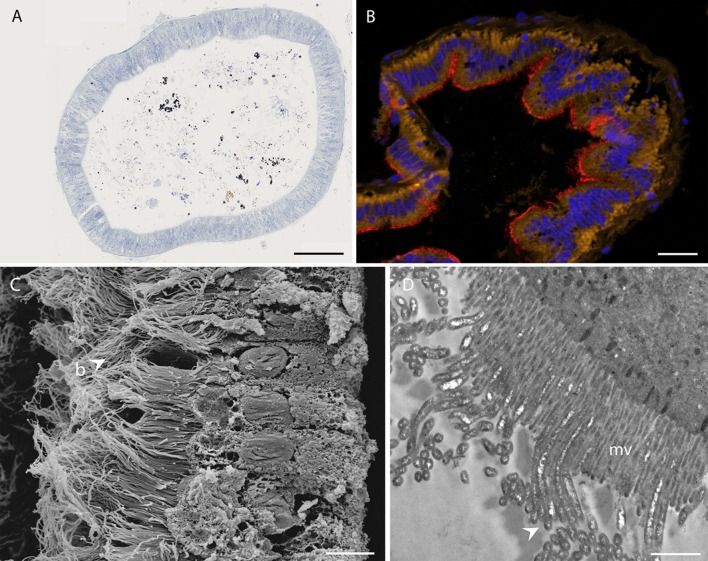
Fig 5. Rimicaris chacei midgut.
A) Photonic observation of a semi-thin section of the digestive tract (hindgut), showing black and brown mineral particles, as well as organic matter stained with toluidine blue. B) FISH of a midgut transversal section stained with DAPI (blue), and hybridized with the Eubacteria general probe Eub338-Cy5 (red) and autofluorescence of intestinal cells (yellow). C) SEM image of a bacterial mat (b) on intestinal wall cells. D) TEM image of filamentous bacteria (arrows) inserted between microvilli (mv) of intestinal cells. Scale bars: A = 10 μm, B = 1 μm, C = 50 μm, D = 10 μm.

