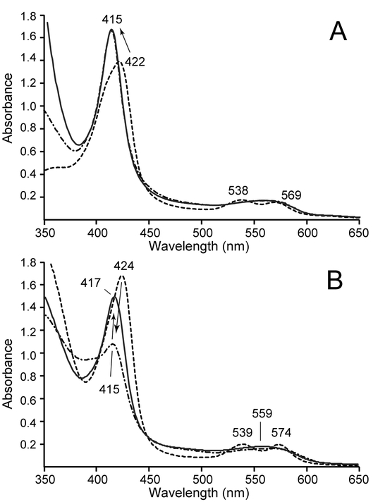
Fig. 8.
Reduction of FeIII–NO GlbN-A and GlbN using heme-free diaphorase and NADPH. (A) The FeIII–NO GlbN-A species (dashed trace) was generated by treatment of FeIII bis–histidine GlbN-A with excess MAHMA-NONOate. Some residual FeIII bis–histidine species is present. Addition of NADPH and heme-free diaphorase resulted in the formation of FeII–NO GlbN-A (dash-dot line), with minimal spectral change upon addition of DT (solid trace). (B) The FeIII–NO GlbN species (dashed trace) was slowly converted to FeII–NO GlbN (dash-dot trace) by incubation with the heme-free diaphorase and NADPH for 70 min. Addition of DT then resulted in the gradual formation of FeII–HNO GlbN (solid trace, ~4 h after DT addition) as evidenced by the increase of the Soret peak to 417 nm.

