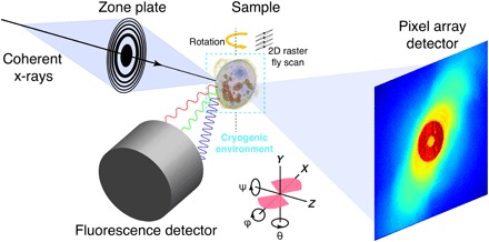
Fig. 1. Experimental schematics for simultaneous x-ray fluorescence and ptychography measurements.

A coherent monochromatic x-ray beam was focused by a Fresnel zone plate into a spot of ~90 nm on a sample. The sample, preserved in the cryogenic environment, was raster fly-scanned in the x-y plane. During the scan, fluorescent signals and diffraction patterns were simultaneously recorded by a fluorescence detector and a pixel array detector, respectively. After finishing a 2D scan, the sample was rotated to a new angle until completing the whole 3D scan. Bottom schematic shows the orientation of 3D reconstructions with respect to experimental setup, where pink regions contain measured projection data between −68° and 56°.
