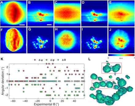
Fig. 4. GENFIRE and FBP reconstruction comparison and angular refinement.

(A to E) FBP reconstructions for ptychography phase contrast, P, S, Ca, and K channels, respectively, projected from the missing wedge direction (i.e., x direction). (F to J) GENFIRE reconstructions of the corresponding volumes shown in (A) to (E), projected from the same missing wedge direction, showing a much better recovery of missing information. Scale bars, 2 μm. a.u., arbitrary units. (K) P channel angular refinement results revealing angular deviations from the recorded tilt axes [ϕ deviation (green), ψ deviation (red), and θ deviation (cyan)]. To see angular orientation with respect to experimental setup, see Fig. 1. (L) Improvements in the P channel 3D reconstruction as a result of angular refinement. Light blue and light green volumes are before and after angular refinement, respectively. Red boxes highlight volumes where angular refinement helped resolve individual acidocalcisomes better.
