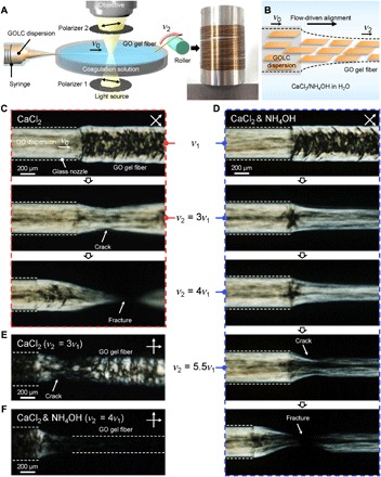
Fig. 1. In situ observation of dynamic self-assembly of GO gel fibers.

(A) Schematic of the experimental set-up: The assembly apparatus was installed on the stage of a POM, and the assembly process was recorded with a high-speed camera focused on the glass nozzle tip. The final GO hydrogel fibers can be reeled on a bobbin as shown in the photo. (B) Schematic illustration showing flow-driven alignment of GO sheets and formation of hydrogel fiber. (C and D) POM snapshots capturing different assembly behaviors of GO gel fibers (C) without and (D) with the addition of NH4OH in the CaCl2 coagulation solution. Snapshots were taken at various rates of the take-up roller (v2). Note that the crossed polarizers were rotated 45° from the fiber axis. (E and F) POM images showing degree of alignment in GO gel fibers obtained at (E) 3v1 in the CaCl2-only coagulation solution and (F) 4v1 in the NH4OH and CaCl2 coagulation solution.
