
Fig. 5. EGFR Inhibition Increases Proteasome-dependent Degradation of MCL1.
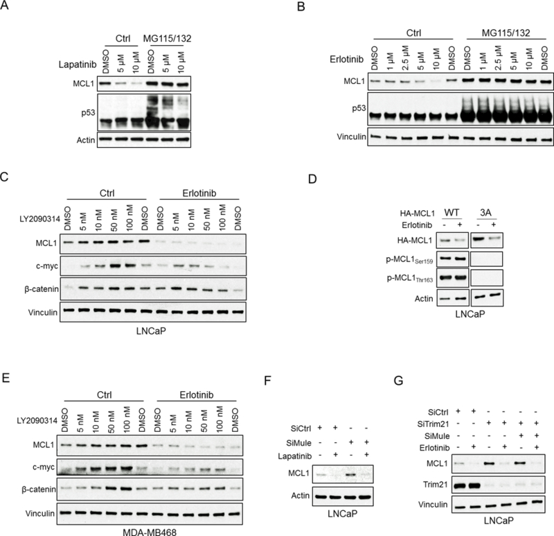
(A and B) LNCaP cells were pretreated with MG115 (10 μM) and MG132 (10 μM) for 30 min, followed by treatment with lapatinib (10 μM) (A) or erlotinib (10 μM) (B) for 4 hours. Efficacy of proteasome block was confirmed by blotting for p53. Results are quantified in Fig. S6B,C. Data shown in A-G are representative of 3 experiments. (C) LNCaP cells were pretreated with GSK3β inhibitor LY2090314 for 1 hour, followed by treatment with erlotinib (10 μM) for 4 hours. Blotting for c-Myc and β-catenin was carried out as a positive control to confirm suppression of GSK3β activity. (D) LNCaP cells were transfected with HA-tagged MCL1 wild-type or 3A (S155A, S159A, T163A) mutant for 24 hours and then treated with DMSO or erlotinib (10 μM) for 4 hours. Lystates were then blotted for HA tagged MCL1, phospho-MCL1 at S159 or T163. (E) MDA-MB468 cells (breast cancer cell line) were pretreated with LY2090314 for 1 hour, followed by treatment with erlotinib (10 μM) for 4 hours. (F) LNCaP cells were transfected with Mule siRNA or non-target siRNA for 48 hours and then treated with DMSO and lapatinib (10 μM) for 4 hours. (G) LNCaP cells were transfected with Trim21 siRNA, double (Trim21 and Mule) siRNA, or non-target siRNA for 48 hours and then treated with DMSO or erlotinib (10 μM) for 4 hours.