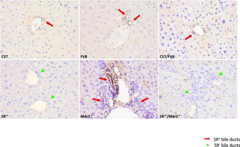
Figure 1.
Expression of SR in liver sections. Immunohistochemistry for SR shows that Mdr2−/− mice have higher immunoreactivity for SR (red arrows depicting bile ducts) compared to WT mice. No immunoreactivity was observed for SR in SR−/− and SR−/−/Mdr2−/− mice (green arrowheads). Original magn., 40×.

