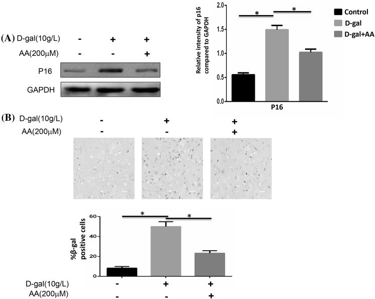
Fig. 3.
Cellular senescence was affected by d-gal and ascorbic acid. BMSCs were stimulated with 10 g/l d-gal and 200 μM ascorbic acid for 48 h. a The protein expressions of p16 were detected by Western blotting. The higher expression of p16 induced by d-gal was reduced when ascorbic acid was added (*P < 0.05). b Quantification of SA-β-gal-positive cells. The total number of SA-β-gal-positive cells among 100 random cells was counted using phase-contrast microscopy. Scale bar = 25 μm. The number of SA-β-gal-positive cells was obviously decreased compared with that in the d-gal treated group (*P < 0.05). Results represent at least three independent experiments

