Fig. 1.
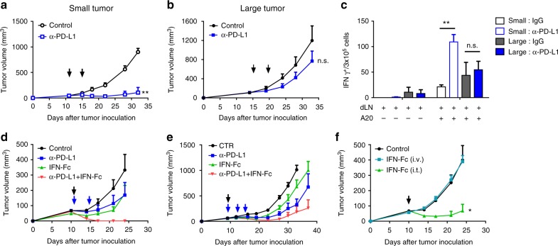
Local delivery of IFNα overcomes resistance to PD-L1 blockade in advanced tumors. a Balb/c mice (n = 3) were inoculated with 3 × 10⁶ A20 cells. Mice bearing early-stage tumors ( < 50 mm3) were treated intraperitoneally (i.p.) with 200 µg of anti-PD-L1 on days 11 and 15. b Mice (n = 4) bearing advanced A20 tumors ( > 100 mm3) were treated with anti-PD-L1 on days 15 and 19. Tumor growth was measured twice per week. c Mice were treated as in a and b. Three days after treatment, cells from draining lymph nodes were isolated and co-cultured with or without irradiated A20 cells for 2 days. The IFNγ ELISPOT assay was performed. d A20 tumor-bearing mice (n = 5) were treated with 200 µg of anti-PD-L1 i.p. on days 11 and 15 and/or 25 µg IFNα-Fc intratumorally (i.t.) on day 11. e C57BL/6 mice (n = 5) were inoculated with 5 × 10⁵ MC38 cells. Mice were treated with 200 µg of anti-PD-L1 i.p. on days 9, 12, and 15, and/or 25 µg of IFNα-Fc i.t. on day 9. The growth curves of MC38 tumors are shown. f A20 tumor-bearing mice (n = 5) were treated i.t. or intravenously (i.v.) with 25 µg of IFNα-Fc on day 11. Black and blue arrows indicate treatment with IFNα-Fc and anti-PD-L1, respectively. Data indicate mean ± SEM and are representative of at least two independent experiments. * p < 0.05; **p < 0.01; n.s. not significant

