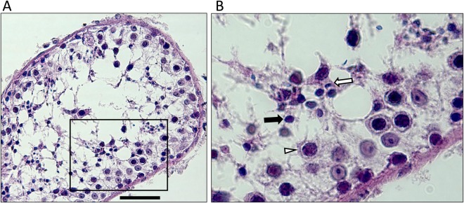
Figure 2.
Histology of testicular tissue of azoospermic male III.3. Formalin-fixed paraffin-embedded tissue was stained with hematoxylin-eosin. Scale bar indicates 50 µm. Severe secondary hypospermatogenesis and spermatocyte stage arrest were observed. Emptied areas filled with building fluid are dispersed inside the tubule. Most cells identified are germ cells from earlier stages, such as spermatocytes (white triangle), and round spermatids (black arrow), with a few elongating spermatids (white arrow).

