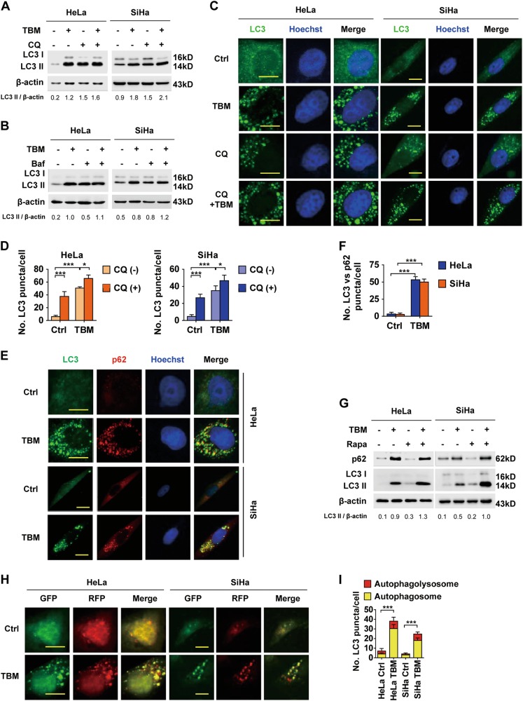
Fig. 5. TBM inhibits autophagic flux in cervical cancer cells.
a HeLa and SiHa cells were treated with TBM in the absence or presence of CQ (10 µM). LC3 was measured with immunoblot. b Cells were treated with TBM in the absence or presence of bafilomycin A1 (Baf, 100 nM). LC3 was measured with immunoblot. c–d HeLa and SiHa cells were treated with TBM in the absence or presence of 10 µM CQ. LC3 puncta formation was measured by immunofluorescence analysis and quantified by ImageJ. e–f Cells were treated with TBM. LC3 and p62 puncta were measured by immunofluorescence analysis and quantified by ImageJ. g HeLa and SiHa cells were treated with TBM in the absence or presence of rapamycin (500 nM). LC3 and p62 expression was measured with immunoblot. The ratio of LC3 II/β-actin was determined with ImageJ software. h–i Cells were transfected with mCherry-GFP-LC3 for 48 h, and treated with TBM for another 24 h. The formation of autophagosome (mCherry-positive; GFP-positive) and autophagolysosome (mCherry-positive; GFP-negative) was examined and quantified by ImageJ. Scale bars: 20 μm. *p < 0.05; ***p < 0.001

