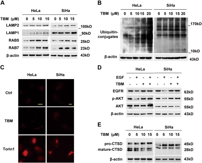
Fig. 6. TBM impairs lysosomal hydrolytic activity in cervical cancer cells.
a Cells were treated with indicated concentrations of TBM for 24 h. The expression of LAMP2, LAMP1, RAB5, and RAB7 in whole cell lysates was determined by immunoblot. b Immunoblot of ubiquitin in HeLa and SiHa cells treated with TBM in indicated concentrations for 24 h. c Autophagolysosomes stained with DQ-BSA in HeLa and SiHa cells treated with 15 μM TBM for 24 h. Accumulation of fluorescent signal indicated the lysosomal proteolysis of DQ-BSA. Torin1 acted as positive control. Scale bar: 10 μM. d HeLa and SiHa cells were starved of serum overnight and treated with EGF (100 nM) in the absence or presence of 15 μM TBM for 2 h. EGFR, AKT and phosphorylation of AKT (Thr 308) were analyzed by immunoblot. e Immunoblot analysis of endogenous CTSD in HeLa and SiHa cells treated with 15 μM TBM for 24 h

