Figure 4.
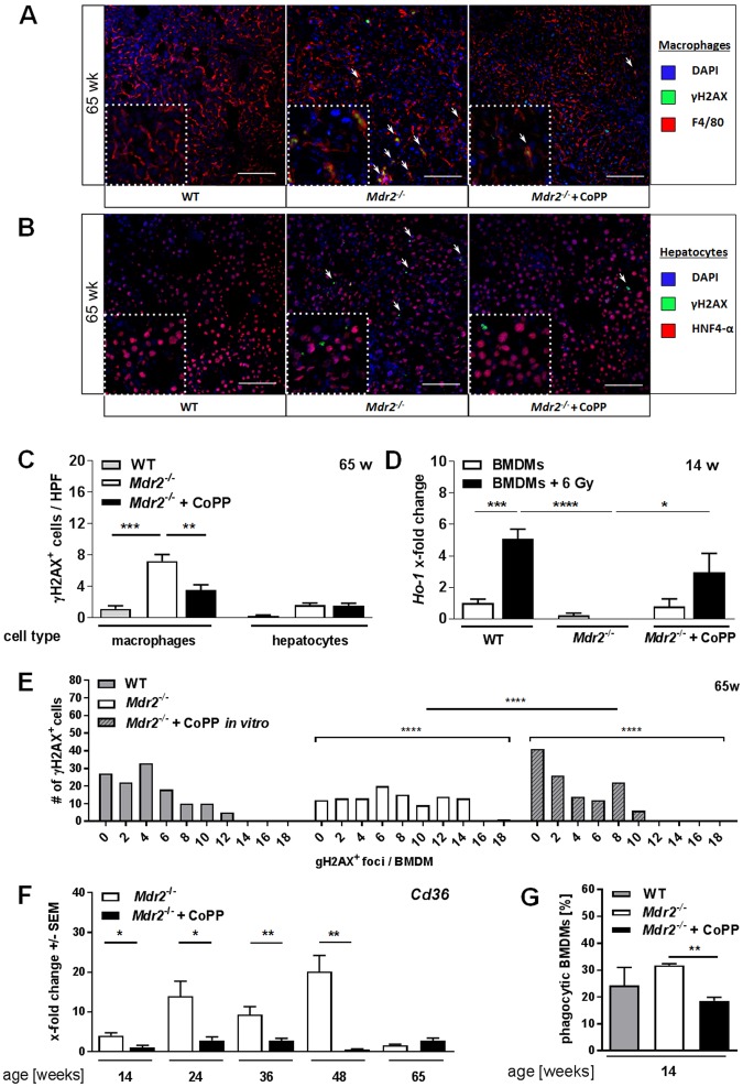
HO-1 induction reduces DNA damage in macrophages in vitro and in vivo. Representative images (20x) of tissue sections of 65-week-old Mdr2−/− mice, treated as described in Suppl. Fig. 1A and WT mice of the same age stained for (A) DAPI, γH2AX, and the macrophage marker F4/80 as well as (B) DAPI, γH2AX, and the hepatocyte marker HNF4-alpha. (C) Quantification of γH2AX+ F4/80+ macrophages and γH2AX+ HNF4-alpha+ hepatocytes in tissue sections described in A (n ≥ 4 HPF/slide). (D) Ho-1 mRNA expression levels of BMDMs derived from 14-week-old WT and Mdr2−/− mice, with or without irradiation, with or without CoPP treatment [10 µg/ml; 24 h prior to irradiation] of one representative experiment (n = 3). (E) Frequency distribution of γH2AX+ foci in BMDMs (65w; WT, Mdr2−/−) with or without CoPP treatment in vitro [10 µg/ml; 24 h]. (F) Hepatic mRNA expression levels of Cd36 determined by quantitative real time RT-PCR in livers of mice described in Suppl. Fig. 1A. (G) Quantification of phagocytic activity of BMDMs derived from 14-week-old animals described in Suppl. Fig. 1A determined by flow cytometry (n ≥ 3; one representative experiment). Data expressed as means ± SEM. *P ≤ 0.05, **P ≤ 0.01, ***P ≤ 0.001, ****P ≤ 0.0001.

