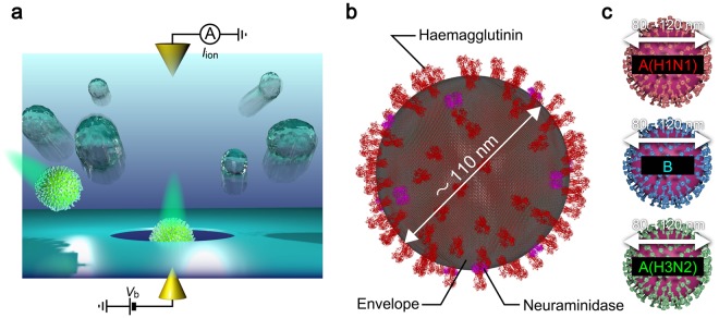
Figure 1.
Single-influenza-virion detections using a solid-state nanopore. (a) Schematic illustration depicting nanopore measurements. Individual influenza virions in chorioallantoic fluid were passed through a Si3N4 nanopore via electrophoresis under the applied voltage Vb and associated resistive pulses were recorded by tracing a temporal change in the cross-membrane ionic current Iion. (b) Influenza virion consisting of a spherical capsid covered with envelope and protein spikes such as haemagglutinin and neuraminidase protruding from the surface. This figure was created using the protein structure of the haemagglutinin32 (https://www.rcsb.org/structure/1ru7) and neuraminidase33 (https://www.rcsb.org/structure/5hun) from the Research Collaboratory for Structural Bioinformatics (RCSB) Protein Data Bank (PDB) website. (c) Three types of influenza viruses employed for nanopore sensing (Top: A(H1N1); middle: B; bottom: A(H3N2)).

