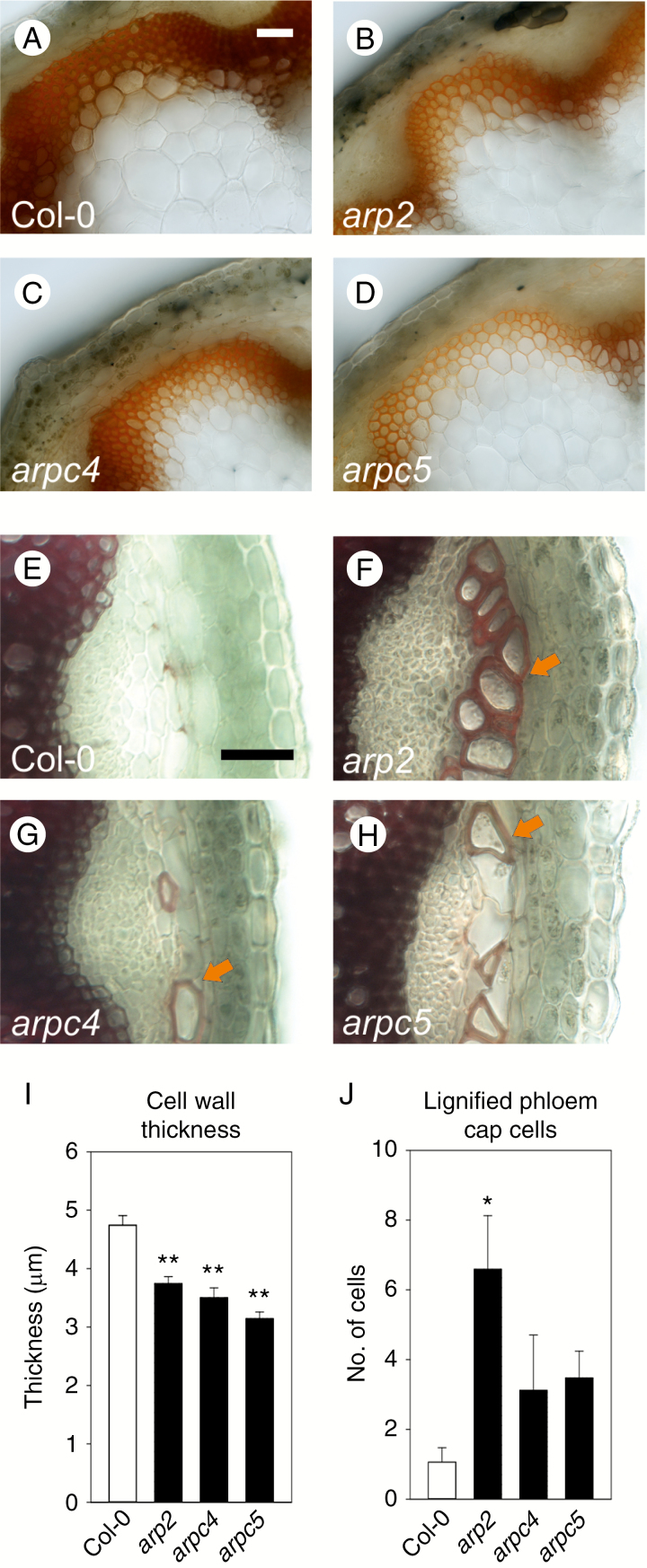
Fig. 4.
Thickness of the cell wall and lignification is altered in ARP2/3 mutants. (A–D) Phloroglucinol HCl staining of transverse sections through the base of the inflorescence stems. Lignification level of interfascicular cells and vascular bundles of arp2 (B), arpc4 (C) and arpc5 (D) mutants is decreased when compared to WT (A). (I) Lignified cell walls in interfascicular tissue shown in A–D are thinner. (E,H,J) In contrast, all mutant plants show increased deposition of lignin in the phloem fibre cells of the phloem cap. Whereas only a few lignified phloem fibre cells per vascular bundle are detectable in WT plants (E, J), a significantly increased number of lignified phloem fibre cells is deposited in the arp2 mutant (F, J). Non-significant increases in the number of lignified phloem fibre cells was detected in arpc4 (G, J) and arpc5 (H, J) mutants. Arrows in F–H indicate lignified phloem fibre cells. Scale bar = 50 µm (A-H). *P <0.05, **P < 0.01.

