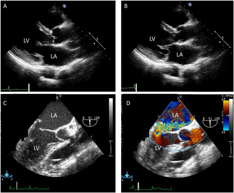
Figure 1.
Transthoracic echocardiography showed MVP at the posterior mitral leaflet [(A) parasternal long-axis view in the diastolic phase and (B) systolic phase]. (C and D) Transesophageal echocardiography showed severe MR from MVP. (D) The color bar shows flow velocity. LA, left atrium; LV, left ventricle.

