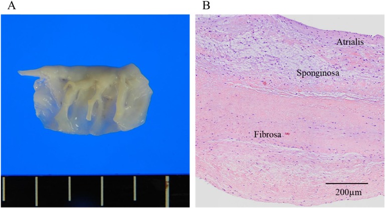
Figure 2.
Histological findings on the mitral valve. (A) Rupture of mitral valve chordae tendineae (P3) was observed. The mitral valve was slightly thickened and soft. (B) Histology (hematoxylin-eosin stain) shows a disorganized fragmentation and tear of collagen and elastin fibers and myxomatous changes. There is no inflammatory cell invasion or granuloma.

