Figure 6.
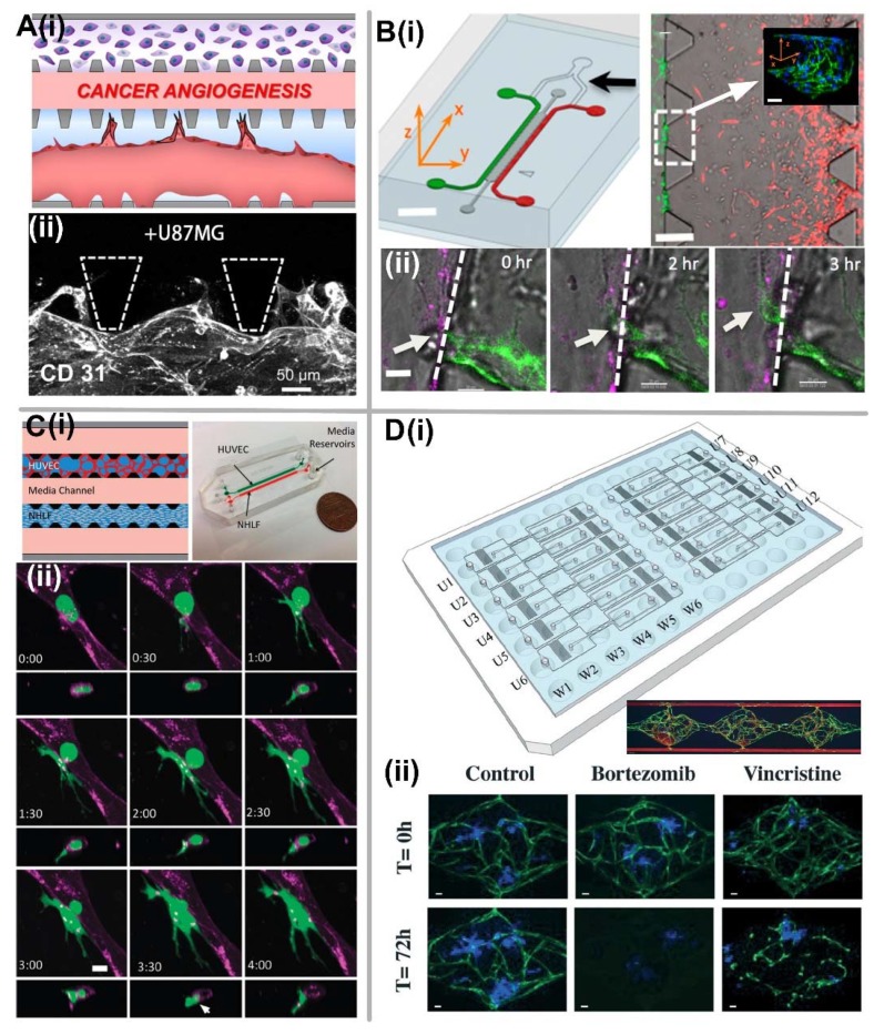
Vascularized tumor-on-a-chip for studying tumor metastasis and anti-cancer drug screening. (A) Tumor angiogenesis. (i) Schematic of chip design for tumor angiogenesis. (ii) 3D confocal image (CD31) shows the angiogenic sprouts stemmed from the microvessel wall grow toward the upper channel stimulated by the pro-angiogenic factors from U87MG cancer cell lines. Adapted by permission from Reference [120], copyright American Institute of Physics 2014. (B) Tumor intravasation. (i) Schematic of microfluidic tumor-vascular interface model, and the immunostaining image showing confluency of the endothelial monolayer on the 3D ECM. Scale bar, 2 mm (left) or 300 μm (right). (ii) Time sequence of a single confocal slice showing a breast carcinoma cell (white arrow) in the process of intravasaton across a HUVEC monolayer. Scale bar, 30 μm. Adapted by permission from Reference [124], copyright National Academy of Sciences, USA 2012. (C) Tumor extravasation. (i) Schematic and prototype of microfluidic device and cell-seeding configuration. (ii) Time-lapse confocal images showing the extravasation process of entrapped MDA-MB-231 (green) from vessel lumen (purple). Scale bar, 10 μm. Adapted by permission from Ref. [126], copyright Royal Society of Chemistry 2013. (D) Large-scale anti-cancer drug screening. (i) Schematic of microfluidic platform design that custom-fitted into a standard 96-well plate format, and the formation of vascularized microtumor tissue inside three tissue chambers. (ii) Drug screening validation with bortezomib and vincristine characterized by the effect on the morphology microvascular network as well as the tumor size. Scale bar, 50 μm. Adapted by permission from Reference [136], copyright Royal Society of Chemistry 2017.

