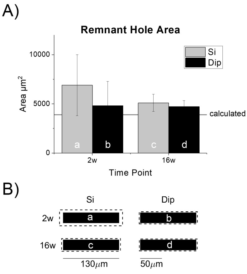
Figure 4.
Characterization of the remnant tissue hole after explantation and ‘dummy’ probe device dimensions. (A) Remnant hole size after probe extraction was consistent (no statistically significant differences) across both implant types. The hole was slightly larger than the theoretical cross-sectional area denoted by the horizontal line; n = 9 (Si-2w), 10 (Si-16w), 10 (dip-2w), 10 (Dip-16w). (B) Mean explanted hole size (dashed line) drawn in relative scale to the actual device dimensions. The letters correspond with the matching bar in the chart shown in (A). The 130 µm scale bar is shown to provide context for the microelectrode dummy probe width. The 50 µm scale bar provides context for the analysis of bucket widths for the histological analysis. Tissue responses can extend several hundred microns away from the tissue-device interface.

