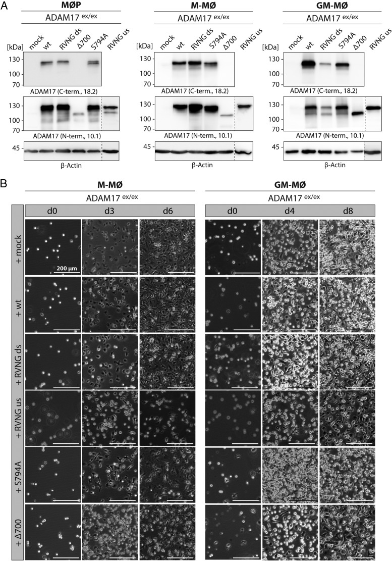
FIGURE 3.
Using the MØP system to study ADAM17 function. (A) MØP with ADAM17-deficient background (ADAM17ex/ex) were stably reconstituted with ADAM17 variants and further differentiated to M-MØ and GM-MØ. By immunoblotting, stable protein expression of each ADAM17 variant was regularly verified using C-terminal (18.2) as well as N-terminal (10.1) Abs. The presented immunoblots are representative for at least four independent differentiations. β-Actin was used as loading control. (B) To ensure equal and complete differentiation of MØP to M-MØ and GM-MØ, cell morphology was monitored by light microscopy for each ADAM17-reconstituted cell line. M-CSF was applied for complete differentiation to M-MØ until day 6 (d6), and representative pictures are shown for day 3 and day 6. Application of GM-CSF had to be continued until day 8 to ensure complete differentiation to GM-MØ, and representative pictures were taken at day 4 and day 8. Undifferentiated MØP are suspension cells, becoming adherent during differentiation growing in characteristic monolayers. Scale bar, 200 μm.

