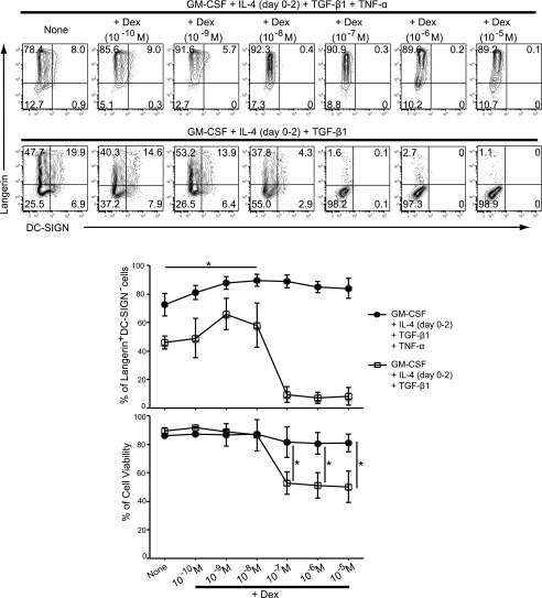
FIGURE 3.
Effect of Dex on the expression of Langerin and DC-SIGN on cells from CD14+ monocytes in the presence of GM-CSF, IL-4, TGF-β1, and TNF-α. Monocytes were cultured in the presence of GM-CSF, IL-4, TGF-β1, and TNF-α with or without different concentrations of Dex (10−10–10−5 M) from initiating the culture for 4 d (upper panels). Monocytes were cultured in the presence of GM-CSF, IL-4, and TGF-β1 with or without different concentrations of Dex from initiating the culture for 4 d (lower panels). Surface expression of Langerin and DC-SIGN was analyzed by flow cytometry. Results shown are representative of four independent experiments, respectively. The percentage of Langerin+DC-SIGN− cells is shown in the middle panel (±SEM). Cell viability was assessed by PI exclusion. The percentage of cell viability from four independent experiments is shown in the bottom panel (±SEM). *p < 0.05, Student t test.

