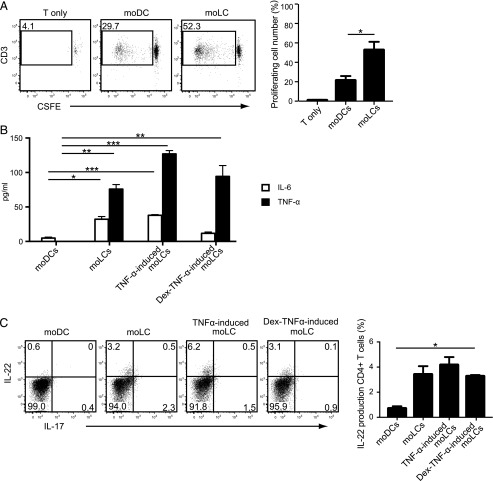
FIGURE 6.
Induction of allogeneic T cell proliferation by coculturing with TNF-α–stimulated activated moLC. (A) CSFE-labeled allogeneic naive CD4+ T cells (1 × 10⁵ cells per well) were cultured with either moDCs or moLCs (2 × 10³ cells per well) for 7 d. The number of proliferated cells is calculated and plotted. (B) The amount of IL-6 and TNF-α in the cell culture supernatants after coculture with moDCs or moLCs and naive allogeneic CD4+ T cells for 7 d was measured by ELISA. (C) Naive allogeneic CD4+ T cells stimulated by moDCs or moLCs were examined for intracellular cytokine expression of IL-22 and IL-17 by flow cytometry. Data are shown as the mean + SEM of results pooled from all three independent experiments. *p < 0.05, **p < 0.01, ***p < 0.001, Student t test.

