FIGURE 1.
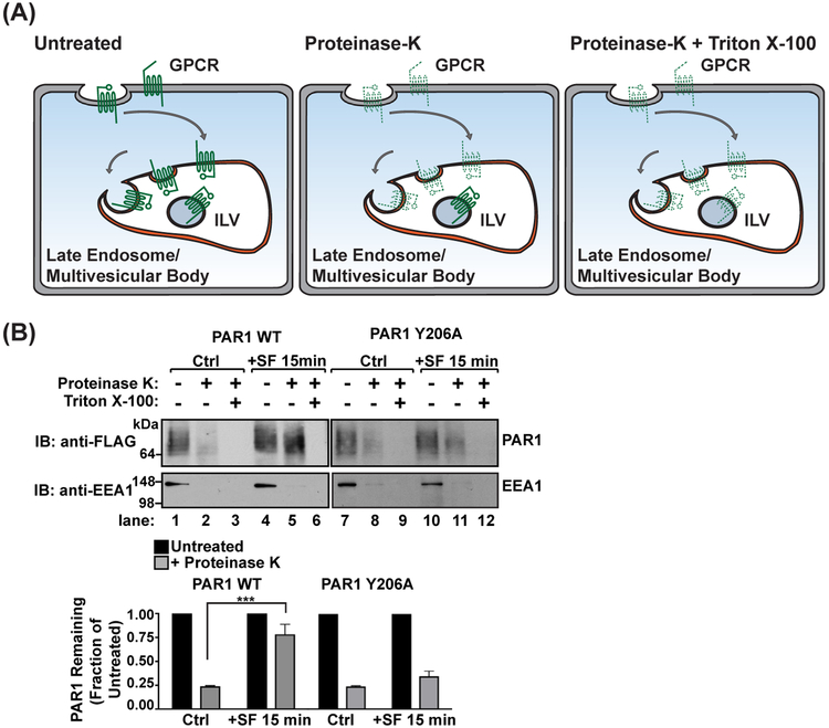
Proteinase-protection endosomal sorting assay. (A) A schematic of proteinase treatment assay with predicted results. Untreated samples represent the total amount of GPCR. Treatment with proteinase K degrades all GPCRs exposed on the plasma membrane or limiting membranes of internal endosomes (dashed lines), whereas GPCRs sorted into intraluminal vesicles (ILVs) are protected from proteinase activity (solid lines). Membranes treated with Triton X-100 detergent acts as a control for proteinase K activity, exposing all receptors to proteinase K degradation. (B) Agonist-stimulated PAR1 sorting into protective endosomal compartments is mediated by the YPXnL motif localized in the second intracellular loop. HeLa cells expressing FLAG-PAR1 WT or FLAG-PAR1 Y206A were stimulated with 100 μM SFLLRN for 15 min. Cells were harvested and treated with digitonin to permeabilize the plasma membrane, then treated with either H2O (untreated), proteinase K, or proteinase K with Triton X-100 detergent. The amount of PAR1 was analyzed by immunoblot and quantified using densitometry (***, p < 0.001, n = 3, 2-way ANOVA)

