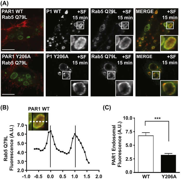
FIGURE 2.
Visualization and quantification of GPCR sorting into expanded endosomes. (A) Agonist-stimulated PAR1 sorting into the lumen of expanded endosomes is mediated by the YPXnL motif. HeLa cells expressing FLAG-PAR1 WT or FLAG-PAR1 Y206A were transfected with Rab5-Q79L-GFP, then surface-labeled with anti-FLAG antibody. Cells were stimulated for 15 min with 100 μM SFLLRN, then fixed and analyzed by confocal microscopy. (B) Line scan analysis of Rab5-Q79L-GFP endosomes. GFP-fluorescence intensity was quantified by line scan (dashed line) and plotted to determine the limiting edges of the endosome (black vertical lines). (C) The PAR1 Y206A mutant is defective for sorting into the lumen of endosomes. PAR1 fluorescence intensity was measured within the limits defined by Rab5-Q79L-GFP (B) and averaged from multiple expanded endosomes (***, p < 0.001, n = 6, Student’s t-test). (See color plate)

