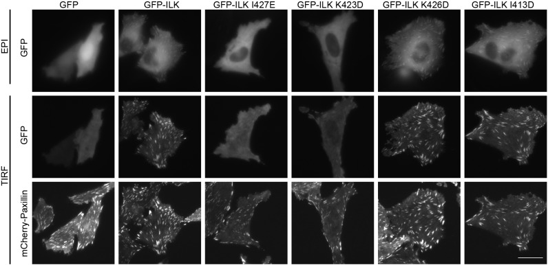
Fig. 6.
GFP–ILK mutants that are impaired in kindlin-2 binding localize poorly to focal adhesions. CHO cells stably expressing mCherry-paxillin were transiently co-transfected with FLAG–α-parvin and either GFP alone, GFP–ILK or one of the GFP–ILK mutants. Six hours after replating on fibronectin-coated glass-bottom dishes, live cells were imaged by epifluorescence (EPI) and/or TIRF microscopy as indicated. Images in each channel were linearly and uniformly adjusted, and cropped for clarity. Scale bar: 20 µm.

