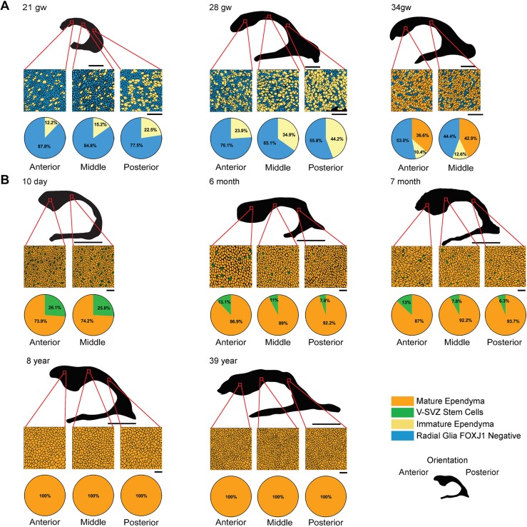
Fig. 2.
Human ependymogenesis proceeds posterior to anterior along lateral ventricle wall during fetal-to-postnatal development and exhibits characteristic pinwheel organization. (A,B) Human fetal (A) and postnatal/adult (B) ependymal cell development was examined at 21 gw, 28 gw, 34 gw, 10 day, 6 months, 7 months, 8 years and 39 years (n=1). Representative schematics of microscope images of a 3391.90 µm2 (fetal) or 13,567.59 µm2 (postnatal) area of the lateral ventricle frontal horn are indicated by red squares on 2D ventricular surface projections (black). Pie charts below schematics indicate percentage of radial glia, immature ependymal cells, V-SVZ stem cells and mature ependymal cells along anterior, middle and posterior regions. Scale bars: 1 cm in A (2D ventricle wall renderings); 5 cm in B (2D ventricle wall renderings); 20 µm in A,B (tissue sections).

