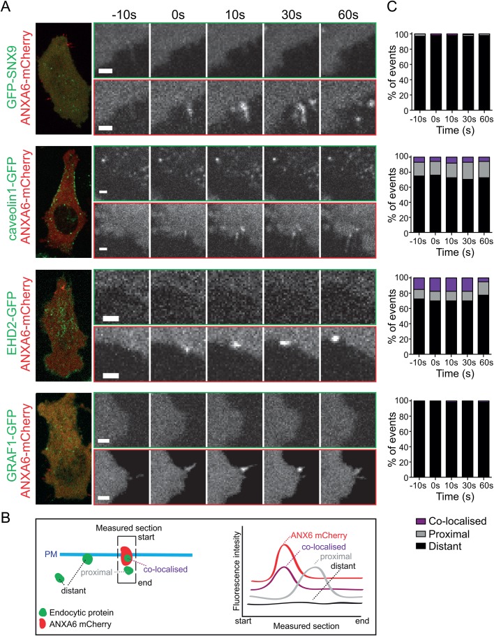
Fig. 3.
Endocytic proteins are not recruited to sites of annexin A6 following LLO addition. (A) Representative images from live-cell confocal spinning disc imaging of ANXA6-mCherry structures and localisation of endocytic proteins to these sites at different time points. To the left is an overview of the cell at time point 0. Movies were taken at 1 fps, time is shown in seconds. Scale bars: 2 μm. Live-cell confocal spinning disc microscopy of GFP-SNX9, caveolin1-GFP, EHD2-GFP and GRAF1-GFP Flp-In TRex cells transfected with ANXA6-mCherry and treated with LLO. (B) Schematic illustration of the plasma membrane (PM) and annexin and endocytic assemblies indicating how the section for measuring intensity of the two channels was drawn, and how the fluorescence intensity was measured and categorised. The section was drawn towards all visible endocytic structures at or in the proximity of annexin A6 structures. The localisation of endocytic proteins to assemblies of ANXA6-mCherry was quantified as; co-localised, proximal (if within 1 μm), and as distant if no correlation was found by analysing the intensity of both GFP and mCherry using a line drawn over the ANXA6-mCherry structures. (C) Quantifications of the localisation of endocytic proteins to the ANXA6-mCherry structures 10 s before the appearance of ANXA6-mCherry (−10 s), at the time of recruitment (0 s) and 10, 30 and 60 s after recruitment. 40–100 events in total were quantified for each endocytic protein.

