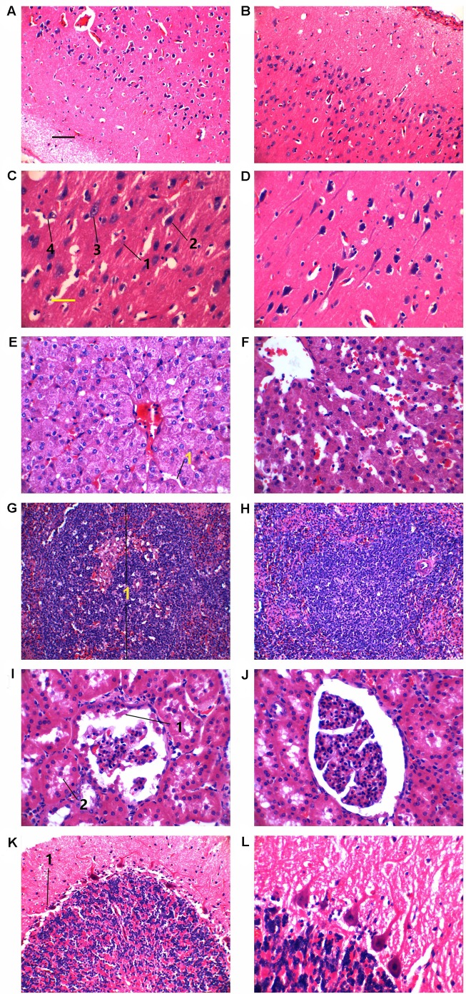
Fig. 5.
Hematoxylin and Eosin staining of tissues from two NAGLU+/− pigs and their two wild-type half sibs at the age of 240 days. (A-D) Cerebra. (E,F) Liver. (G,H) Spleen. (I,J) Kidney. (K,L) Cerebella. In C: 1, shortened pyramidal cells; 2, nuclei in neurons appeared shrunk and stained darker; 3, vacuole formation; 4, mild microgliosis. In E: 1, narrow hepatic sinusoid. In G: 1, one splenic corpuscle. In I: 1, hyaline droplets; 2, albumin leakage. In K: 1, one purkinje neuron. Left panels, NAGLU+/− pigs. Right panels, wild-type half sibs. Black scale bar for A,B,G,H: 50 µm. Yellow scale bar for C-F,I-L: 25 µm.

