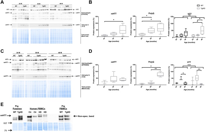
Fig. 5.
TgHD minipig PBMCs do not express mHTT. (A-D) Western blot analyses of individual minipigs and subsequent quantification revealed that mHTT and endogenous HTT were expressed in a tissue- and age-specific manner in the frontal cortex (A,B) (mHTT: TgHD 24 vs 48 months, P=0.04; PolyQ: TgHD 24 vs 48 months, P=0.02; TgHD 24 vs 36 months, P=0.02; HTT: TgHD 24 vs 48 months, P=0.03; TgHD 36 vs 48 months, P=0.02; ANOVA WT vs TgHD, P=0.004) and putamen (C,D) (PolyQ: TgHD 24 vs 48 months, P=0.04; TgHD 36 vs 48 months, P=0.004; HTT: WT 36 vs 48 months, P=0.04; TgHD 36 vs 48 months, P=0.009; ANOVA WT vs TgHD, P=0.001) in TgHD minipigs, and confirmed the absence of mHTT and PolyQ in WT animals. (E) mHTT was not expressed in minipig TgHD PBMCs. Representative western blot of mHTT in PBMCs from HD patients (HD) and controls (Ctr), and PBMCs from WT and TgHD minipigs, as indicated. Three distinct antibodies were used to identify mHTT and PolyQ fragments and endogenous HTT protein (see Materials and Methods). Mann–Whitney test and ANOVA, *P<0.05, **P<0.01, ***P<0.001. Sample sizes: (A,B) mHTT and PolyQ: TgHD 24 months, n=4; TgHD 36 months, n=5; TgHD 48 months, n=6; HTT: WT 24 months, n=3; WT 36 months, n=4; WT 48 months, n=6; TgHD 24 months, n=4; TgHD 36 months, n=5; TgHD 48 months, n=5; (C,D) mHTT and PolyQ: TgHD 24 months, n=3; TgHD 36 months, n=6; TgHD 48 months, n=5; HTT: WT 24 months, n=2; 36 months, n=3; 48 months, n=5; TgHD 24 months, n=3; 36 months: n=6, 48 months, n=5.

