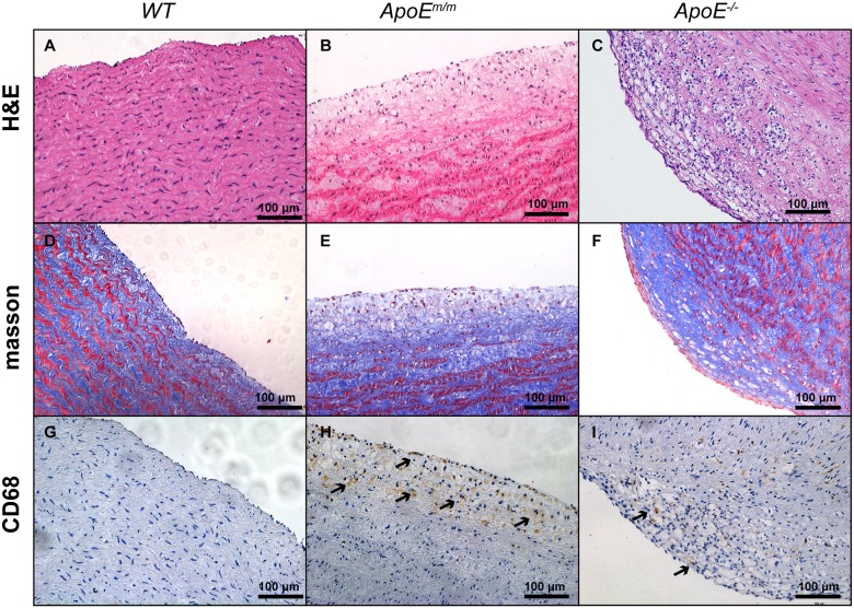
Fig. 6.
Representative histological images of aorta from WT, ApoEm/m and ApoE−/− pigs. No visible atherosclerosis was seen in the aorta from WT pigs (A,D,G). ApoEm/m pigs had pathological fatty streak of the aorta (B,E,H). ApoE−/− pigs showed progressive atherosclerotic lesions in the luminal part of the arterial intima (C,F,I).

