Fig. 3.
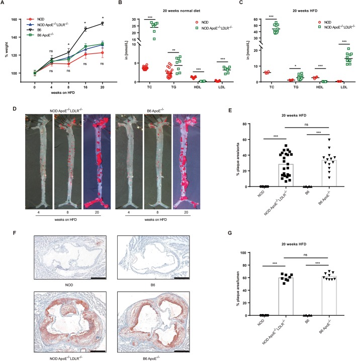
Hyperlipidemia and atherosclerosis in double-knockout NOD Apoe−/−Ldlr−/− mice. (A) Kinetics of body weight of NOD Apoe−/−Ldlr−/− and B6 Apoe−/− mice fed a HFD for 20 weeks, with NOD and B6 wild-type controls. For each time point, at least three animals were analyzed for each genotype (n=3-55). (B) Blood cholesterol (TC), triglyceride (TG), high-density lipoprotein (HDL) and low-density lipoprotein (LDL) concentrations of mice on normal diet for 20 weeks analyzed in two independent experiments involving NOD Apoe−/−Ldlr−/− and NOD control mice (n=8-19). ‘in [mmol/L]’, the levels of TC, TG, LDL and HDL were measured in mmol/l. (C) Blood TC, TG, HDL and LDL concentrations of mice on a HFD for 20 weeks analyzed in two independent experiments involving NOD Apoe−/−Ldlr−/− and NOD mice (n=4-11). (D) Representative images of Oil-Red-O-stained aortas of mice on a HFD for 4, 8 and 20 weeks (en face assay). (E) Quantitative analysis of en face lesion area of NOD Apoe−/−Ldlr−/− and B6 Apoe−/− mice fed a HFD for 20 weeks, with NOD and B6 wild-type control mice [NOD, n=6 (male=3, female=3); NOD Apoe−/−Ldlr−/−, n=23 (male=10, female=13); B6, n=6 (male=3, female=3); B6 Apoe−/−, n=13 (male=4, female=9)]. Data were collected from two independent experiments with the same animals used in D (n=6-23). (F) Four representative microscopy images of aortic root sections from NOD Apoe−/−Ldlr−/− and B6 Apoe−/− mice placed on a HFD for 20 weeks, with controls of NOD and B6 wild-type mice. Scale bars: 400 μm. (G) Quantitation of plaque area relative to the area of the aortic lumen from F [NOD, n=6 (male=3, female=3); NOD Apoe−/−Ldlr−/−, n=8 (male=5, female=3); B6, n=6 (male=3, female=3); B6 Apoe−/−, n=9 (male=4, female=5)] (n=6-9). Statistics by two-tailed, unpaired Student's t-test: *P<0.05, **P<0.01, ***P<0.001, ns, not significant.

