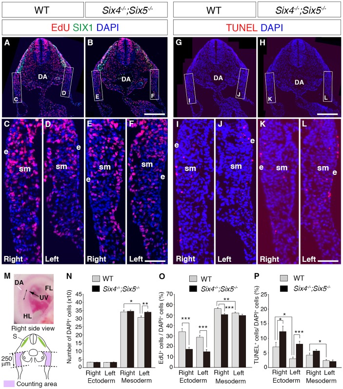
Fig. 5.
Altered cell proliferation rate in the PAW of Six4−/−;Six5−/− embryos. (A-F) Distribution of EdU-positive cells after EdU incorporation for 30 min in the PAW of wild-type (A,C,D) and Six4−/−;Six5−/− (B,E,F) embryos at E10.5. The SIX1 signal, which is used as a marker for muscle progenitor cells, is observed in wild-type and Six4−/−;Six5−/− embryos (A,B). (G-L) Distribution of TUNEL-positive apoptotic cells in the PAW of wild-type (G,I,J) and Six4−/−;Six5−/− (H,K,L) embryos at E10.5. (M) A right-side view of an E10.5 mouse embryo. A double-headed arrow (↔) indicates the anterior-posterior level of the analyzed sections. The illustration shows the counting area of EdU- and TUNEL-positive cells in the PAW (pink). (N) The number of cells within the counting area. The number of mesodermal cells in the left side is smaller than that in the right side of the PAW. (O) The ratio of proliferating cells in the PAW. The numbers of EdU-positive cells in the ectodermal and mesodermal cells of the right and left PAW, and the ratio and total cell number were calculated. In wild-type embryos, the ratio of proliferating cells in mesodermal regions of the PAW is higher in the right side than in the left side. In Six4−/−;Six5−/− embryos, the ratio of proliferating cells in the ectodermal region of the PAW is lower in both right and left sides of the PAW than that in the wild-type embryos, whereas the ratio of mesodermal cells is lower in the right side but similar in the left side compared with wild-type embryos. (P) The ratio of TUNEL-positive cells. In wild-type embryos, the ratio of TUNEL-positive cells in the ectodermal and mesodermal regions of the PAW are larger in the right side than that in the left side. The ratio of TUNEL-positive cells in Six4−/−;Six5−/− embryos is larger than that of wild-type embryos in the right and left ectoderm of the PAW. Error bars show the standard error. *P<0.05, **P<0.01, ***P<0.001, Student's t-test, n=3. DA, dorsal aorta; e, ectoderm; FL, forelimb; HL, hindlimb; S, somite; sm, somatic mesoderm; UV, umbilical vein. Scale bars: 250 μm in A,B,G,H; 50 μm in C-F,I-L.

