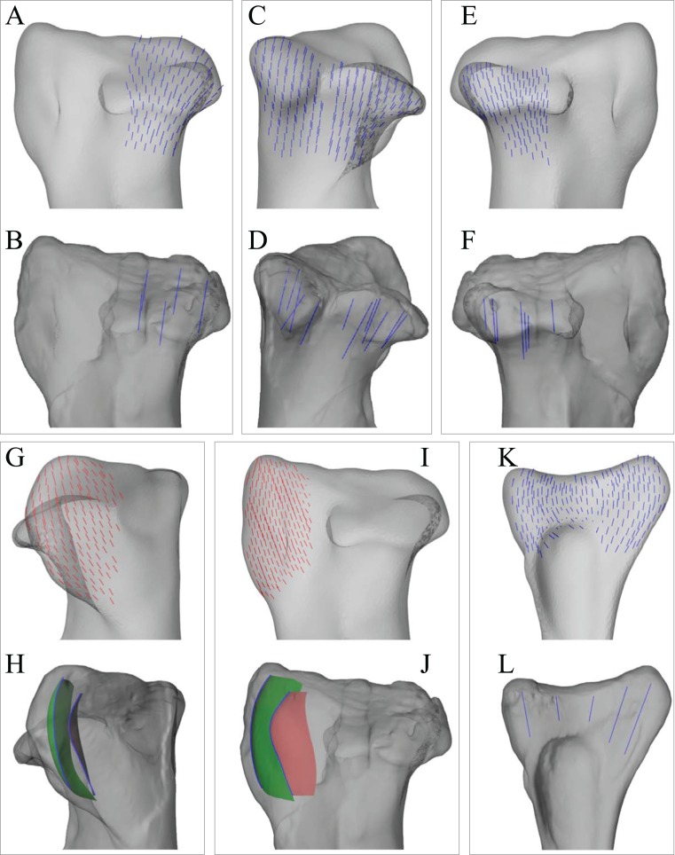
Figure 8. Principal stress trajectories for the tibia and fibula in the solution posture for Daspletosaurus, compared with observed cancellous bone fabric.
(A) Vector field of σ3 in the medial tibial condyle, shown as a 3-D slice through the middle of the condyle and parallel to the sagittal plane, in medial view. (B) Observed cancellous bone architecture in the medial tibial condyle of Allosaurus and tyrannosaurids (cf. Part I), in the same view as (A). (C) Vector field of σ3 in the medial and lateral tibial condyles, shown as 3-D slices through the middle of the condyles and parallel to the coronal plane, in posterior view. (D) Observed cancellous bone architecture in the medial and lateral tibial condyles of Allosaurus and tyrannosaurids (cf. Part I), in the same view as (C). (E) Vector field of σ3 in the lateral tibial condyle, shown as a 3-D slice through the middle of the condyle and parallel to the sagittal plane, in lateral view. (F) Observed cancellous bone architecture in the lateral tibial condyle of Allosaurus and tyrannosaurids (cf. Part I), in the same view as (E). (G) Vector field of σ1 in the cnemial crest, shown as a 3-D slice parallel to the coronal plane, in anterior view. (H) Observed cancellous bone architecture in cnemial crest of Allosaurus and tyrannosaurids (cf. Part I), sectioned in the plane of the crest, shown in the same view as (G); blue section lines illustrate primary architectural direction. (I) Vector field of σ1 in the cnemial crest, shown as a 3-D slice parallel to the sagittal plane, in medial view. (J) Observed cancellous bone architecture in cnemial crest of Allosaurus and tyrannosaurids (cf. Part I), sectioned in the plane of the crest, shown in the same view as (I). (K) Vector field of σ3 in the medial aspect of the fibular head, in medial view. (L) Observed cancellous bone architecture in the fibular head of Allosaurus and tyrannosaurids (cf. Part I), in the same view as (K).

