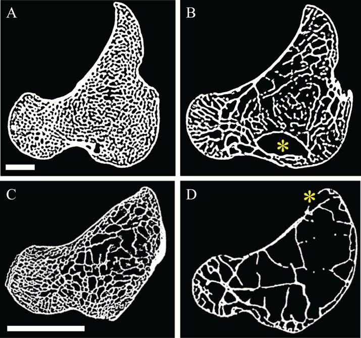
Figure 14. Pneumatization modulates trabecular spacing in the femur of both large and small birds.
This is illustrated here with processed CT scan slices located approximately midway through the femoral head in the axial plane. (A) A marrow-filled femur of a southern cassowary, Casuarius casuarius (QMO 30105), with mean trabecular spacing = 0.638 mm. (B) A pneumatized femur of an emu, Dromaius novaehollandiae (QMO 16140) with mean trabecular spacing = 1.128 mm. (C) A marrow-filled femur of a chicken, Gallus gallus (PJB coll.), with mean trabecular spacing = 0.320 mm. (D) A pneumatized femur of an Australian brush turkey, Alectura lathami (PJB coll.), with mean trabecular spacing = 0.999 mm. Reported trabecular spacing values were calculated (for illustrative purposes) for the femoral head using the BoneJ 1.3.11 plugin for ImageJ (Doube et al., 2010). (A) and (B) are shown at the same scale, as are (C) and (D). Scale bars are 10 mm, and yellow asterisks denote pneumatopores.

