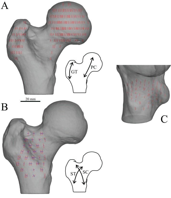
Figure 15. The main architectural features of cancellous bone in the human proximal femur.
(A) Vector field of u1 in the head, inferior neck and greater trochanter regions, plotted on a translucent rendering of the external bone geometry; view is in the coronal plane. Schematic inset illustrates close correspondence with the principal compressive (PC) and greater trochanter (GT) groups of previous studies. (B) Vector field of u1 (red) and u2 (blue) in the middle of the metaphysis, viewed in the coronal plane. Schematic inset illustrates close correspondence with the secondary compressive (SC) and secondary tensile (ST) groups of previous studies. Note that both u1 and u2 are largely parallel to the coronal plane. (C) Vector field of u1 in the distal metaphysis and lesser trochanter (in oblique proximomedial view), which is largely parallel to the bone’s long-axis. In this and all subsequent illustrations of fabric vector fields, the length of each fabric vector is proportional to its corresponding fabric eigenvalue. Additionally, all images are of bones from the right side of the body.

