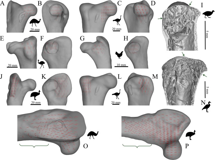
Figure 16. The main architectural features of cancellous bone in the proximal femur of birds.
(A–D) Vector field of u1 in the femoral head and inferior neck of an emu, Dromaius novaehollandiae (QMO 16140; A, B), and ostrich, Struthio camelus (MV R.2385; C, D). (E–H) Vector field of u1 under the facies antitrochanterica of a greater rhea, Rhea americana (QMO 23517; E, F), and chicken, Gallus gallus (PJB coll.; G, H). (I) Isosurface rendering of cancellous bone under the facies antitrochanterica of a dusky moorhen, Gallinula tenebrosa (PJB coll., between arrows), sectioned in the sagittal plane. (J–M) Vector field of u1 in the trochanteric crest of a southern cassowary, Casuarius casuarius (QMO 30105; J, K), and Struthio camelus (MV R.2385, L, M). (N) Isosurface rendering of cancellous bone in the trochanteric crest of a magpie goose, Anseranus semipalmata (QMO 29529, between arrows), sectioned in the sagittal plane. (O, P) Vector field of u1 throughout the entire proximal femur of Dromaius novaehollandiae (QMO 16140, O) and Casuarius casuarius (QMO 30105, P), which illustrates the increasing obliquity and disorganization of vectors in the distal metaphysis and transition to the diaphysis, shown in regions with braces. (A, C, J and L) are anterior views; (B and D) are medial views; (E and G) are posterior views; (F, H, K and M) are lateral views; (O) is an oblique anterolateral view; (P) is an oblique anteromedial view. For reference, silhouettes of the animals depicted are provided in this figure and those that follow.

