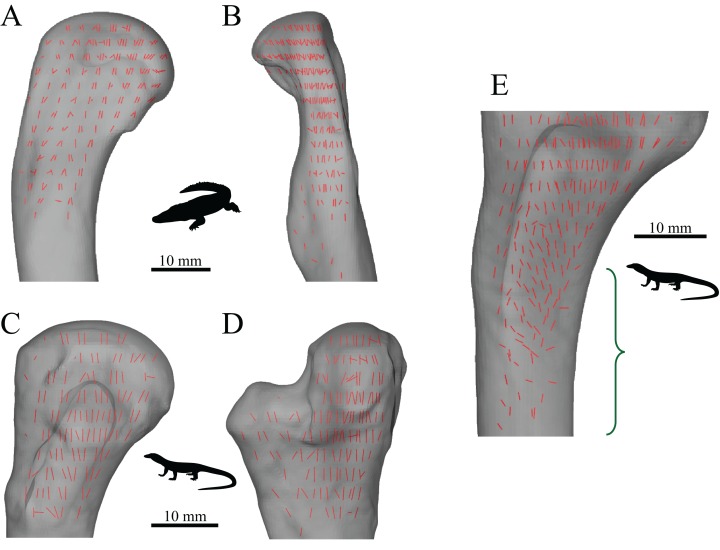
Figure 17. The main architectural features of cancellous bone in the proximal femur of extant sprawling reptiles.
(A, B) Vector field of u1 in the proximal femur of a freshwater crocodile, Crocodylus johnstoni (QMJ 47916). (C, D) Vector field of u1 in the proximal femur of a Spencer’s goanna, Varanus spenceri (QMJ 84416). (E) Vector field of u1 throughout the proximal femur of a Komodo dragon, Varanus komodoensis (AM R.106933), which illustrates the increasing obliquity and disorganization of vectors in the distal metaphysis and transition to the diaphysis, shown in region with braces. (A and C) are anterior views (‘dorsal view’ of herpetologists); (B and D) are lateral views (‘posterior view’ of herpetologists); (E) is an oblique anterolateral view. For clarity, the vectors of u1 in the fourth trochanter are not visible in (A, C and E).

