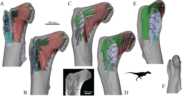
Figure 19. The main architectural features of cancellous bone in the proximal femur of both Allosaurus and the tyrannosaurids.
These are illustrated here with a 3D geometric model of the observed architecture, mapped to the femur of Daspletosaurus torosus (TMP 2001.036.0001). (A–E) Five progressive rotations of the bone, in 30° increments, from anteromedial to anterolateral views (C is a purely anterior view). (F) The observed orientation of the dominant tract of cancellous bone in the femoral head (blue) has a gentle anterior inclination; bone shown in medial view. For explanation of the features and colour coding, refer to the main text. Inset below C is a CT slice through the proximal femur of Tyrannosaurus rex (MOR 1128), parallel to the coronal plane and through the middle of the femoral head. This illustrates the very characteristic tract of cancellous bone that extends from the base of the femoral neck up towards the apex of the head, highly comparable to the tract present in humans (cf. Fig. 4A).

