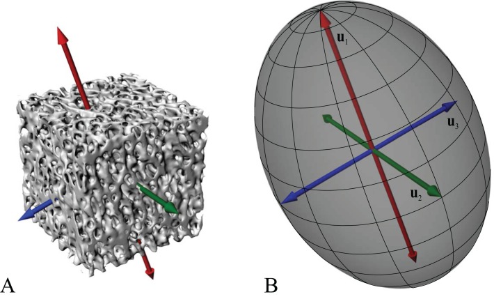
Figure 2. Cancellous bone fabric as represented by its principal architectural directions.
(A) A cube of cancellous bone of side length 5.33 mm, from the proximal femur of a freshwater crocodile, Crocodylus johnstoni, with the principal directions of the bone’s architecture superimposed. As an orthotropic material, cancellous bone fabric is completely described by three principal directions. (B) The fabric ellipsoid representation for this cube of cancellous bone is derived from the vectors that describe the principal architectural directions. The ellipsoid’s major, semimajor and minor axes are given by the primary (u1), secondary (u2) and tertiary (u3) directions of the cancellous bone architecture, which correspond to the eigenvectors of the fabric tensor. The relative lengths of each axis depend on the relative magnitudes of the principal directions, which correspond to the eigenvalues of the fabric tensor. The degree of anisotropy (DA) describes the extent to which the trabeculae are aligned within a sample, and is given as the relative magnitude of the primary and tertiary eigenvalues (i.e. DA = e1/e3); in this instance DA = 1.44. The cancellous bone geometry was derived via micro-computed X-ray tomographic scanning (Siemens Inveon, 80 kV, 500 µA, 900 ms exposure, 53.3 µm isotropic resolution) and 3D visualization (Mimics 17.0, Materialise NV, Belgium). The material directions were calculated using the mean intercept length method as implemented in the software Quant3D 2.3 (see Ketcham & Ryan, 2004).

